Abdominopelvic ultrasound: interest, procedure and management of the examination
1. How does the act take place?
The abdominopelvic ultrasound takes place in a radiology office or in a hospital, in a medical imaging department. It lasts an average of 15 minutes.
The patient will generally be asked to present on an empty stomach (4 hours minimum), which includes the absence of tobacco and coffee. This preparation is necessary to correctly visualize the gallbladder and to allow better exploration of the digestive tract. On the other hand, you should not stop your usual treatments.
In some cases, it will be recommended to come with a full bladder (having drunk 1 liter of water during the hour preceding the examination), for a better visualization of the wall of the bladder, the ovaries, and the digestive tract. Indeed, many intestinal loops are in the lower abdomen and contain air. These digestive gases hamper the search for lesions and this parameter is corrected.
Abdominal or pelvic ultrasound is performed by a radiologist. Some midwives are competent to perform ultrasounds during the course of an uncomplicated pregnancy.
Before the examination, the doctor asks the patient about his general condition, his history and his possible treatments.
The examination takes place in a room where the light is dimmed, to facilitate the reading of the images on the screen, and includes several stages.
The patient is lying on an examination table, on his back, and undressed at the level of the abdomen.
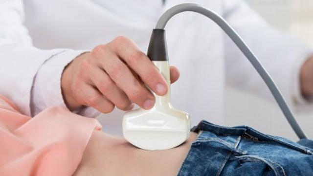
The doctor applies a skin gel to the skin of the stomach, to promote contact between the probe and the skin, which is essential for the proper transmission of ultrasound.
He then places the probe in contact with the skin and moves it over the stomach, sometimes exerting slight pressure, in order to visualize the different organs. The doctor may sometimes ask the patient to stop breathing for a few moments, thus freeing up certain areas of the liver and spleen, and obtaining higher quality images by limiting the natural movements associated with breathing.
Ultrasound has no real adverse effects. The patient may feel an impression of cold given by the gel. When the doctor has to exert pressure to analyze certain organs, it can be uncomfortable, especially if the area was already painful. Finally, having a full bladder can create an unpleasant feeling.
When the examination must be completed by an endovaginal or endorectal route, it is necessary to empty the bladder beforehand, to remove any vaginal tampon and to report an allergy to latex (the probe being covered with a disposable protection latex).
To perform a transvaginal ultrasound, the position is similar to that of a gynecological examination, on the back, with the knees bent and the soles of the feet on the examination table. The practitioner introduces a probe provided for this purpose into the vagina, wrapped in sterile protection, covered with a lubricant, then moves it slightly in order to observe the uterus and ovaries. This technique cannot be used on a minor or a woman who has never had sexual intercourse.
In the case of a transrectal ultrasound, the practitioner lubricates the anal canal so that the sensation is the least unpleasant possible when inserting the probe. The procedure is then the same as for an endovaginal route, with a probe covered with a sterile single-use protection, which will be placed in different positions to observe the organs. This technique can also be used to guide the performance of prostate biopsies.
During the examination, the ultrasound echoes returned by the organs are picked up by the probe, which transmits them to a computer which converts them into moving images, visible on a screen, in real time.
The radiologist then records so-called reference images, on which he wishes to measure certain parameters (the size of an organ, the flow within the vessels, the presence of an anomaly, the growth parameters of the fetus, etc.).

Listen to our latest episode of the CAREcast as our prevention team discusses how to define hookups, establish boun… https://t.co/8O8nO7iDdR
— UC Merced CARE Wed Jul 21 18:38:49 +0000 2021
Once the abdominopelvic ultrasound is completed, the doctor interprets the results and explains them to the patient. He writes an examination report, generally containing the key images he has produced, which he also sends to the prescribing doctor or the treating doctor.
Depending on the results of the ultrasound, he may advise the performance of additional examinations, to complete the assessment (for example a scanner or a biological assessment).
2. Why have an abdominal ultrasound?
An abdominal ultrasound can be prescribed for different reasons in order to make a diagnosis. The main indication is the exploration of abdominal pain. It is also recommended in the event of the discovery of jaundice, renal symptoms (renal colic, presence of blood in the urine), or abnormalities in the biological assessment concerning in particular hepatic or renal functions.
It makes it possible to diagnose various pathologies on the various solid organs of the abdomen and pelvis, including stones or dilation in the gallbladder, liver damage, pancreatitis, pathologies of the spleen such as spenomegaly, aneurysm, thrombosis, masses in the kidney, kidney stones, enlarged prostate, ovarian cysts or endometriosis.
It also makes it possible to analyze certain digestive structures. In an emergency context, it makes it possible to look for appendicitis, in particular in young adults and especially in children.
On the pediatric level, it should be noted that ultrasound is the key imaging examination in the face of vomiting in children. Depending on the age, the radiologist can make the diagnosis of pyloric stenosis or intussusception, which cause vomiting in the very young.
Ultrasound examination of the digestive tract may show the presence of thickening of the intestinal walls of the small or large intestine. Such images are often non-specific and may sometimes evoke an infectious cause (colitis), Crohn's disease in a young person or diverticular disease or even a tumor in an older person.
During pregnancy, abdominal ultrasound can monitor the proper growth of the fetus and detect certain morphological abnormalities. In conventional pregnancy follow-up, three ultrasounds are thus recommended.
It can, in certain specialized centers, be used to guide a biopsy or a puncture, carried out by the radiologist or by a surgeon.
Finally, it is used in the monitoring of certain pathologies: monitoring of cancer, monitoring of kidney or liver transplantation
3. What convalescence?
None ! Ultrasound is a painless, non-invasive, non-irradiating examination with no side effects.
The images and the report of the ultrasound are given to the patient by the radiologist just after the examination.
However, depending on the results observed, other examinations may possibly be prescribed to clarify a diagnosis (scanner, MRI, complementary biology).
Read also :
⋙ Jaundice: causes, symptoms and treatments for jaundice
⋙ Irritable bowel syndrome: reduce abdominal pain
⋙ Renal colic: diagnosis, examinations and treatments
Effects of palm oil on health: what are the dangers?
GO
Eudaemonism: all about this philosophy of happiness
GO
Vaccination obligation in Lourdes: the employee of a dialysis center dismissed
GO
Charlotte, student midwife: "We are very quickly in autonomy"
GO